
Bone Fractures and Healing : Expert care from an Orthopedician
What is a Bone Fracture?
A bone fracture is a disruption in the integrity of a bone, typically resulting from trauma, high-impact forces, or conditions such as osteoporosis that compromise bone strength, leading to a crack or complete break in the bone tissue. It can range from minor hairline cracks to complete breaks that displace bone fragments, potentially disrupting normal function and requiring medical intervention for proper healing. Fractures are classified by their type, location, and severity, and they typically present with symptoms such as pain, swelling, bruising, and limited mobility.
Fracture v/s Break
A bone fracture and a bone break are often used interchangeably to describe an injury where the bone's integrity is compromised; however, "fracture" is the more precise medical term that highlights the specific nature of the injury, including various types such as simple, compound, or stress fractures, and provides a clearer understanding for diagnosis and treatment. While "break" is a more colloquial, everyday expression used to describe the same injury in simpler terms.
Fracture v/s Bruise
A bone fracture is a break or crack in the bone that often causes significant pain, swelling, and sometimes visible deformity, requiring immobilization or surgical intervention for healing. In contrast, a bone bruise, also known as a bone contusion, involves bleeding and swelling within the bone tissue without a complete break, often resulting from trauma and causing deep, aching pain that may be less apparent externally but can be detected through advanced imaging like MRI. While fractures typically demand more immediate and invasive treatment, bone bruises tend to heal gradually with rest and symptom management, highlighting their distinct impacts on bone integrity and recovery processes.
Fracture v/s Sprain
A bone fracture involves a break or crack in the bone structure, often causing intense pain, swelling, bruising, and difficulty moving the affected area, and typically requires immobilization or surgical intervention for healing. In contrast, a sprain is an injury to ligaments—the tissues connecting bones at joints—usually resulting from overstretching or tearing, characterized by pain, swelling, bruising, and joint instability, but generally does not involve a broken bone and often heals with rest, ice, compression, and elevation.
Different Types of Bone Fractures
Fractures are classified based on their pattern, cause, and location, with each aspect providing vital information for diagnosis and treatment. A transverse fracture is characterized by a horizontal break that traverses the bone, typically resulting from a direct blow or trauma, whereas an oblique fracture occurs at an angled or diagonal line across the bone, often caused by twisting forces. In contrast, a comminuted fracture involves the bone splintering into several fragments, usually due to high-impact injuries or severe trauma, making the treatment more complex and requiring careful stabilization to ensure proper healing. The cause of the fracture—such as a high-impact trauma leading to a direct blow or a stress fracture from repetitive activity—also influences its classification, as does the specific body part affected, like a clavicle, femur, or wrist, since different bones have unique structural and healing properties. This comprehensive classification helps an orthopedician in Guntur to tailor treatment strategies to ensure optimal recovery.
Articles List
- How Sitting Too Long Damages Your Back Expert Orthopedic Surgeon Advice
- Understanding Pneumonia: Causes, Symptoms, And Risk Factors
- Healthy Lifestyle for Strong Bones tips from the best Orthopedician in Guntur
- Sports Injuries: Types, Symptoms, Causes, and Treatment Options in Guntur
- How to Prevent Joint Pain Expert Health Care Tips for Healthy Bones and Muscles
- Lung Disease causes, symptoms and when to See a Pulmonologist
- Myths and Facts About Knee & Shoulder Arthroscopy, You Should Know
- Best Ways to Keep Your Lungs Healthy and Strong
- Bone Fractures and Healing : Expert care from an Orthopedician
Based on pattern
Fractures are classified based on their pattern, with some involving a single straight-line break such as oblique, transverse, and longitudinal fractures, while others involve more complex patterns like greenstick, comminuted, segmental, and spiral fractures, which do not follow a single straight line and often indicate different mechanisms of injury and severity.
Based on cause
Fractures can be classified based on their underlying causes, such as stress fractures resulting from repetitive mechanical stress leading to tiny cracks, avulsion fractures caused by a sudden force pulling a fragment of bone away from its main structure, and buckle fractures, which occur when the bone compresses and buckles upon itself, often seen in children due to their flexible bones.
Based on location
Fractures can be categorized based on their anatomical location, impacting various regions such as the upper extremities—including the clavicle, shoulder, humerus, elbow, ribs, and facial bones— as well as the hands and wrists, with specific types like Barton, Chauffeur, Colles, Smith, Scaphoid, and metacarpal fractures. The lower limbs also have distinct fracture types affecting the pelvis, acetabulum, hip, femur, patella, growth plates, tibia, and fibula, while the feet and ankle region includes injuries such as calcaneal stress fractures, fifth metatarsal fractures, Jones fractures, Lisfranc injuries, talus fractures, trimalleolar, and pilon fractures. This classification helps in diagnosis and tailoring appropriate treatment strategies for each specific injury. Each location-specific fracture can sometimes present as multiple types, especially in high-impact trauma, like a severe fall causing a comminuted tibia fracture, which involves the shin bone breaking into multiple pieces. Notably, fractures in the feet and ankles, such as Jones or Lisfranc fractures, are particularly prone to complications like nonunion, where the bone fails to heal properly. Emphasis on precise diagnosis and customized treatment plans based on the fracture's exact location is crucial for optimizing healing outcomes and minimizing complications, ensuring that each patient receives care specifically aligned with their injury's unique characteristics.
Open v/s closed fracture
Your healthcare provider will determine whether your fracture is open or closed; an open fracture, also called a compound fracture, occurs when the broken bone protrudes through the skin, increasing the risk of infection and often requiring more extensive treatment, while a closed fracture involves a break in the bone without breaking the skin, though it remains a serious injury that needs proper care for healing.
Displaced v/s non-displaced fracture
Your healthcare professional will classify your fracture as either displaced or non-displaced based on the alignment of the broken bone fragments, with displaced indicating misalignment and non-displaced meaning the fragments remain in proper position. A displaced fracture occurs when the bone fragments shift significantly, creating a noticeable gap or misalignment, often necessitating surgical intervention from an orthopedic surgeon in Guntur to realign and stabilize the bone. Conversely, a non-displaced fracture means the bone is broken but the fragments remain in proper alignment, typically allowing for conservative treatment such as casting without surgery.
Symptoms of Bone Fractures
Symptoms of bone fractures often encompass severe pain, noticeable swelling, and tenderness around the affected area, along with difficulty moving the limb or joint as usual. Additional signs include bruising or discoloration due to bleeding under the skin, and sometimes a visible deformity or unusual bump that indicates a misaligned bone, which can be mistaken for swelling or swelling-related changes but signals a break.
Causes of Bone Fractures
Bone fractures typically result from significant trauma, such as car accidents, falls, or sports injuries, where a force strong enough to break the bone is applied. However, fractures can also occur without a direct traumatic event, often due to repetitive stress from activities like running or overuse from repetitive motions, which may lead to stress fractures or overuse syndromes, especially in the hands and arms. Individuals with osteoporosis face a heightened risk of fractures, as this condition weakens bones and accounts for over a million fractures annually, emphasizing the importance of bone health and preventive measures.
Diagnosis and Tests
To diagnose bone fractures, orthopedic hospitals in Guntur utilize various imaging modalities, including X-rays, which are the primary tool to confirm fractures and assess the extent of bone damage. MRI scans offer a comprehensive view of both bones and surrounding tissues such as cartilage and ligaments, providing detailed soft tissue information, especially in complex injuries. CT scans provide highly detailed cross-sectional images, making them useful for evaluating intricate fractures or those obscured on X-rays. Bone scans are particularly helpful for detecting fractures that are not visible on X-rays, as they identify areas of increased bone activity, though they require multiple visits and are more time-consuming.
Treatment and Management
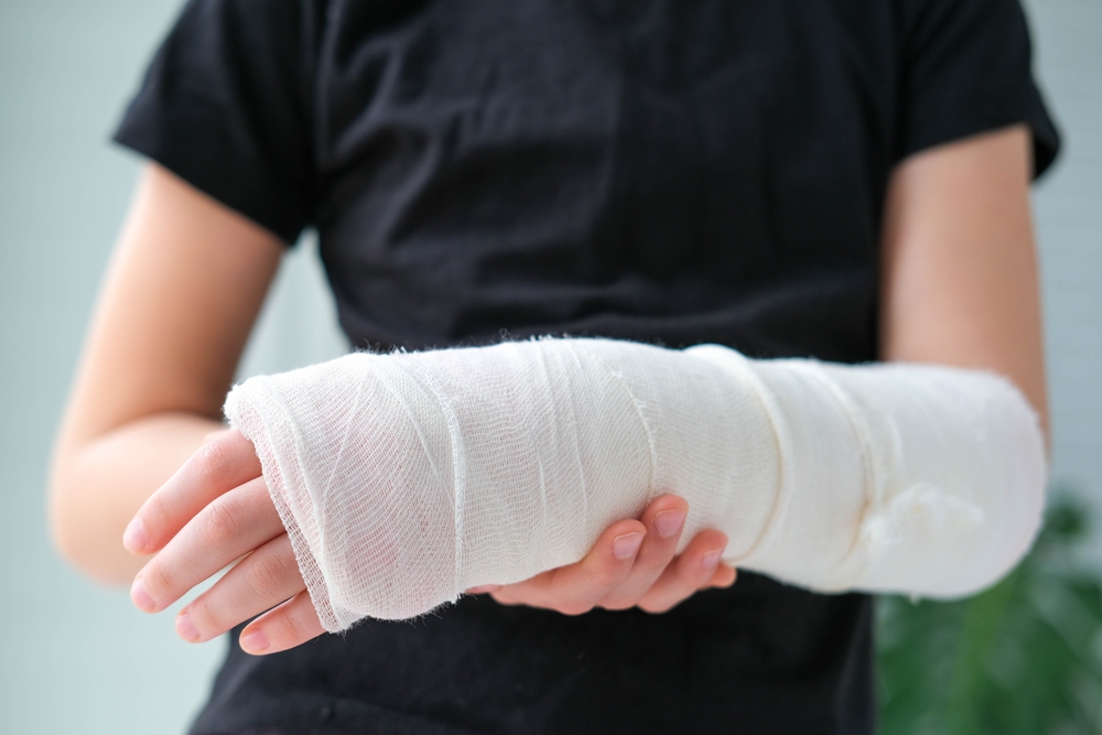
Treatment for a fracture varies based on its type, cause, and severity, with options including immobilization through splints or casts for minor, non-displaced fractures, typically lasting 3-8 weeks and requiring follow-up X-rays; more complex or displaced fractures often necessitate a closed reduction, a non-surgical procedure where a healthcare provider realigns the bones externally under local anesthesia, sedation, or general anesthesia, followed by immobilization with a splint or cast to ensure proper healing. Certain bone fractures necessitate surgical intervention, with the specific approach tailored to the fracture's type and severity; techniques such as internal fixation with plates and screws, external fixation devices, or minimally invasive procedures like percutaneous pinning may be employed to ensure proper alignment and healing.
Internal fixation
Internal fixation is a surgical procedure where your surgeon realigns broken bones and stabilizes them using metal hardware to facilitate proper healing and growth. Techniques include the insertion of rods that run longitudinally through the bone, plates attached with screws to hold fracture fragments, and pins or wires for smaller or more delicate bone structures, often used alongside rods or plates. While some individuals live with these implants permanently, others may require additional surgeries to remove them once healing is complete, depending on their specific situation and the type of hardware used.
External fixation
External fixation is a surgical technique where your surgeon inserts screws into the bone on both sides of a fracture and connects them externally with a brace or frame, providing stabilization while the bone begins healing. Typically used as a temporary measure, this method allows the fracture to stabilize before considering internal fixation options, helping to manage complex or open fractures and reducing the risk of further injury or infection during the healing process.
Arthroplasty
When a joint such as the shoulder, elbow, or knee sustains a fracture, surgical intervention may involve arthroplasty, where the damaged joint is excised and substituted with a prosthetic component. This artificial joint, crafted from durable materials like metal, ceramic, or high-grade plastic, is designed to closely mimic the appearance and function of a natural joint, enabling restored mobility and improved quality of life.
Bone grafting
Bone grafting may be necessary if your fracture is significantly displaced or if your bone isn’t healing properly, involving the addition of extra bone tissue to aid in proper rejoining. Surgeons typically perform internal fixation to stabilize the fractured segments during healing, which can involve using bone grafts sourced from your own body (often the pelvis), a donor, or synthetic materials. Following the procedure, your bone will require immobilization with devices such as a splint, cast, brace, or sling to ensure proper healing before resuming normal activity.
Complications of Bone Fracture Treatment
Complications following fracture surgery can be serious and multifaceted, including acute compartment syndrome (ACS), where increased pressure within muscle compartments impairs blood flow, risking irreversible tissue damage; malunion, where bones heal improperly aligned, potentially impairing function; nonunion, where the bone fails to heal completely, necessitating further intervention; osteomyelitis, an infection of the bone that is more likely with open fractures exposing the bone to bacteria; and collateral internal injuries, such as damage to surrounding muscles, nerves, blood vessels, tendons, and ligaments, which can complicate recovery and require additional treatment.
Medication for Bone Fracture
Over-the-counter NSAIDs such as aspirin and ibuprofen are effective for pain relief but can increase the risk of bleeding, ulcer formation, gastrointestinal discomfort, and bowel issues, especially following surgery; therefore, your surgeon will recommend appropriate medications to manage pain safely and minimize these potential complications.
Healing Time
The healing time for a bone fracture varies based on several factors such as the cause, the specific bone affected, the nature of the fracture, the treatment used, and any additional injuries sustained. Typically, minor fractures may allow for movement within a few weeks, while more severe breaks can take a year or longer to fully recover. It's important to follow your healthcare provider’s guidance throughout the healing process and to seek immediate medical attention if you experience persistent or intense pain that does not improve.
Outlook
While many individuals fully recover from broken bones and return to their usual activities, certain fractures—especially those accompanied by additional injuries—may lead to lasting effects. It's essential to consult with orthopedic doctors in Guntur before resuming sports or strenuous activities to ensure proper healing and avoid complications during your recovery process.
General Safety Tips
To minimize injury risks, always buckle your seatbelt and wear appropriate protective gear during sports and activities. Keep your home and workspace organized to prevent trips and falls and use step stools or proper tools instead of standing on furniture when reaching for items. Maintain a balanced diet and regular exercise routine to promote strong bones and consult your healthcare provider about getting a bone density test if you're over 50 or have a family history of osteoporosis. If you experience walking difficulties or have a higher fall risk, utilize assistive devices like canes or walkers for added stability and safety.
From the Doctor’s Desk
Seeking urgent medical attention is crucial if you experience trauma with signs such as severe pain, restricted movement, visible deformity, bone protrusion, swelling, or simultaneous bruising, as these may indicate a fracture. While fractures don’t directly cause fevers, the presence of a fever, warmth, or redness around the injury suggests potential infection, necessitating immediate medical evaluation to prevent serious complications. While fractures are a frequent injury, they can be quite frightening to endure. If you experience a broken bone, consult Dr. Chaitanya Ghanta a leading orthopedic doctor at Sree Sankalpa to understand the healing process and what to anticipate. Fortunately, most individuals recover fully and can resume their normal activities without lasting effects. It's important to be patient with your recovery, even though it can be challenging, because taking adequate time allows your bone to heal properly and reduces the risk of re-injury.